Chapter 7. Pregnancy
! 700
Chapter Authors & Contributors
- Nova Panebianco, MD
- Robert M. Mayall, BHSc, PhD
- Jonathan Wallace, MD, RDMS
- Adam Guthrie, BKin, MMgmt
7.1 Introduction
The goal of early obstetric ultrasound is to rule in the presence of an intrauterine pregnancy.
A yolk sac is the first definitive sign of pregnancy.
Complications of 1st trimester pregnancy are common emergency department (ED) problems. Miscarriage occurs in 20-25% and ectopic pregnancy in 1.5-2% of pregnancies.[1] Studies have shown that ED physicians can accurately and rapidly use bedside ultrasound to detect an intrauterine pregnancy (IUP).[2][3][4]
The goal of early obstetric ultrasound is to rule in the presence of an IUP. Because heterotopic pregnancy is extremely rare in patients who have not undergone reproductive assistance, ectopic pregnancy can be ruled out with the presence of an intrauterine pregnancy with a negative predictive value approaching 100%.[5] This assumption may not hold true in women who have taken fertility drugs or received in vitro fertilization.
In most ED settings, a definitive IUP is determined when a yolk sac or fetal pole (+/- a heartbeat) is identified within an intrauterine gestational sac on either transabdominal or transvaginal ultrasound (#Image 7.1 and #Image 7.2).
Image 7.1
Transabdominal Yolk Sac
! 700
Image 7.2
Transvaginal Yolk Sac
! 700
Further investigation may be warranted when bedside ultrasound does not conclusively identify an IUP, a patient has had assisted conception (even in the presence of an IUP because of the possibility of a heterotopic pregnancy), and when an IUP is noted on US but with significant pain or fluid within the pelvis (#Image 7.3, #Movie 7.1 and #Movie 7.2).
Image 7.3
Free Fluid
! 700
Movie 7.1
Empty Uterus
##### Movie 7.2 **IUP with Free Fluid**7.2 Getting Started
- A bedside ultrasound should be considered in any patient with early pregnancy and symptoms/signs of potential ectopic pregnancy (e.g. pelvic pain, vaginal bleeding, hypotension, etc.).
- The Pouch of Douglas is the most dependent area in the female pelvis.
- Transabdominal scanning is best performed with a full bladder.
- Transvaginal scanning should be performed with an empty bladder.
- Image the uterus in long and transverse planes.
A bedside ultrasound should be considered in any patient with early pregnancy and symptoms/signs of potential ectopic pregnancy (e.g. pelvic pain, vaginal bleeding, hypotension, etc.).
For transabdominal ultrasound (TAUS), a curved array general abdominal probe (2-5 MHz) is preferable, though a phased array probe will suffice. If no IUP is identified on transabdominal scanning, transvaginal ultrasound (TVUS) is performed with the endocavitary probe (5-8 MHz) (#Gallery 7.1).
Gallery 7.1
Probes
[[Pasted image 20251227151223.png]] | Curvilinear probe
[[Pasted image 20251227151241.png]] | Transvaginal probe
Relevant Anatomy
The uterus is pear-shaped, located between the urinary bladder and the colon. In the majority of women, the uterus is anteroflexed; however, in about 10% of women, it is retroflexed (#Gallery 7.2 and #Movie 7.3).
Gallery 7.2
Pelvic Anatomy
[[Pasted image 20251227151419.png]] | Normal anatomy
[[Pasted image 20251227151436.png]] | Anteroverted/anteroflexed uterus
[[Pasted image 20251227151459.png]] | Retroverted/retroflexed uterus
Movie 7.3
Transvaginal Uterus Long Axis
The Pouch of Douglas (recto-uterine pouch) is the most dependent area in the female pelvis. Fallopian tubes are rarely seen on ultrasound except when pathologic (e.g. hydrosalpinx/tubo-ovarian abscess) (#Image 7.4). Ovaries are typically 2x3cm, and lie lateral to the body of the uterus. They are situated antero-medial to the internal iliac vessels (#Movie 7.4 and #Movie 7.5).
Image 7.4
Hydrosalpinx
! 700
Movie 7.4
Ovary
Movie 7.5
Left Ovarian Cyst
### Technique The technique for early obstetric ultrasound will now be described (#Gallery 7.3). The initial exam should be transabdominal. This gives a big-picture view of the female pelvis. If pertinent structures are not adequately visualized, perform a TVUS. For both exams, the patient should be supine and appropriately draped. ##### Gallery 7.3 **Technique** ```captiongallery [[Pasted image 20251227151715.png]] | Transvaginal sagittal/long axis [[Pasted image 20251227151735.png]] | Transabdominal full bladder [[Pasted image 20251227151757.png]] | Transvaginal coronal/short axis [[Pasted image 20251227151818.png]] | Transabdominal long axis [[Pasted image 20251227151842.png]] | Endometrial stripe [[Pasted image 20251227151859.png]] | Pouch of Douglas ```Transabdominal is best performed with a full bladder. In the longitudinal plane, the indicator is to the head. Track the vaginal stripe to the cervix, and the endometrial stripe through the uterus. The pelvic structures are often not midline and you must scan from right to left until the best plane showing these structures in longitudinal section is found. Increase the depth to visualize the posterior uterus and Pouch of Douglas. Obtain a video and/or still images of the pertinent structures. Rotate the transducer 90 degrees counter-clockwise (marker to 9 o’clock) into the transverse plane. Again, scan through the entire uterus, cervix to fundus. Do not focus on the gestation (if visualized) until the uterus has been systematically evaluated in both long and transverse planes. Bowel gas, body habitus, or an empty bladder may limit your view. Constant, firm pressure may initiate peristalsis or displace bowel. If possible, wait for the bladder to fill by hydrating the patient. The following video demonstrates transabdominal ultrasound to rule in IUP (#Movie 7.6).
Movie 7.6
One Minute Ultrasound IUP Demo
Transvaginal is more difficult than TAUS because of orientation. Efficient TVUS is performed immediately following the pelvic examination, obviating the need to reposition the patient and find a chaperone twice. The pelvic exam will also inform the sonographer regarding location and orientation of the pelvic structures. An empty bladder is essential for the exam. Place gel on the endocavitary probe, cover with a protective barrier (condom, glove, or commercial probe cover), and then apply sterile gel to the outside (#Movie 7.7).
Movie 7.7
One Minute Ultrasound Transvaginal Demo
Structures closest to the probe will appear at the top of the US viewing screen. When the probe indicator is facing up toward the ceiling, a long axis view of the uterus is obtained. When the probe indicator is facing 9 o’clock (toward the patient’s right), a transverse view of the uterus is obtained.
For a longitudinal image (#Gallery 7.4), insert the probe along the anterior vaginal canal, indicator toward ceiling. Once the long image of the uterus is found (in many patients, it may be off of the midline), make a mental note of the location and plane of the transducer. Systematically scan from cervix to fundus (pointing the US beam toward the ceiling to see the fundus, and toward the floor to see the cervix in the anteverted uterus), right to left, through the entire uterus. For the transverse plane, rotate the probe counterclockwise so that the indicator faces the patient’s right (9 o’clock), keeping the uterus in view at all times. Scan through the entire uterus from cervix to fundus, checking for normal and abnormal findings. A full bladder, retroverted uterus, or fibroids may limit your view (#Gallery 7.4).
Gallery 7.4
Transvaginal Anatomy
[[Pasted image 20251227152114.png]] | Transvaginal coronal/short axis
[[Pasted image 20251227152136.png]] | Transvaginal transverse
[[Pasted image 20251227152214.png]] | Transvaginal sagittal/long axis
[[Pasted image 20251227152214.png]] | Transvaginal long
[[Pasted image 20251227152230.png]] | Transvaginal long axis
[[Pasted image 20251227152251.png]] | Large uterine mass - fibroid
The adnexa may be scanned with TAUS, though imaging is typically better with TVUS (#Movie 7.8 and #Image 7.5). Scan the right and left adnexa in transverse plane to identify the ovaries and note any abnormal findings. Ovaries are typically located at the level of the body of the uterus, anterior and medial to the internal iliac vessels (#Movie 7.4 and #Movie 7.5). However, they may be anywhere from the midline in the Pouch of Douglas, to high in the pelvis and quite lateral.
Ovarian parenchyma is slightly hypoechoic compared to myometrium. Ovarian follicles are fluid filled cystic structures and appear anechoic when gain settings are correct. Ovarian parenchyma, ringed by cysts, has a sonographic appearance of “chocolate chip cookies.” Ovaries are often difficult to identify on TAUS. Increase depth and lower transducer frequency to locate the ovaries and surrounding structures. Pelvic vessels and bowel are often misidentified as ovaries. Apply colour flow and wait for peristalsis if in doubt.
Movie 7.8
Right Adnexa with Blood and Ectopic
##### Image 7.5 **Right Adnexa** 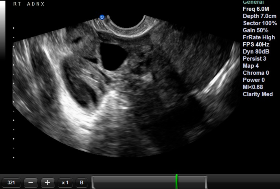7.3 Ultrasound Findings
- The double decidual sign appears around a beta-hCG of 1,000-2,000.
- Serum beta-hCG level alone cannot differentiate intrauterine from ectopic pregnancy.
- Consider a FAST exam in an unstable pregnant patient.
- A pseudogestational sac can be confused for a gestational sac in an ectopic pregnancy.
- If there is greater than a 50% subchorionic hemorrhage, then the prognosis for the gestation is poor.
Normal US Findings
The goal of this study is to identify a definitive IUP. The double decidual sign/sac (DDS) forms as the early implantation enlarges, deforming the surrounding endometrium, resulting in two roughly concentric, thick, hyperechoic endometrial rings (#Gallery 7.5, image 1). The DDS is the only sonographic finding that is consistently related to quantitative β-hCG (Qβ-hCG) levels. It is reliably seen by TVUS when the beta-hCG is between 1,000-2,000. Typical transvaginal “discriminatory zone” values, or the beta-hCG level in which one would expect to see evidence of an IUP, range in the literature from 1,500 to 3,000 mIU/mL and have largely been developed according to radiology-performed ultrasonography in outpatient settings.6 The beta-hCG quantitative level should not determine whether or not to do the scan, as >50% of women with ectopic pregnancy have a beta-hCG level less than 2,000mIU.7 Additionally, in a study by Wang et al., when an emergency physician–performed pelvic ultrasonography result was indeterminate for IUP, serum β-hCG level alone did not differentiate intrauterine and ectopic pregnancy.[6]
Since the interpretation of the DDS is subjective, many take a more conservative approach and do not consider it a definitive sign of IUP. The presence of a yolk sac (YS) or fetal pole (FP) within an intrauterine gestational sac is considered the first definitive sign of an IUP. A yolk sac is an echogenic circle/ring within an intrauterine gestational sac (#Gallery 7.5). A fetal pole may range from nonspecific echogenic material to a well-formed fetus, depending on the gestational age. The fetal heart rate (FHR) may often be identified near the base of the YS before a definite FP is seen.
The fetal heart rate is most commonly measured in M-mode. It can be measured by spectral Doppler; however, the time-averaged acoustic intensity delivered to the fetus is higher with this modality than with M-mode and is not recommended for more than 4-5 beats.[7] Place the M-mode line across the beating heart to obtain a FHR tracing. With most machines, the FHR is calculated by placing calipers from a position of one beat to the equivalent location of the next beat (#Gallery 7.5, images 5 and 6). Normal range is 100-180 BPM.
Gallery 7.5
Normal Ultrasound Findings
[[Pasted image 20251227152653.png]] | Double decidual sign
[[Pasted image 20251227152712.png]] | Transabdominal yolk sac
[[Pasted image 20251227160420.png]] | Yolk sac and fetal pole
[[Pasted image 20251227160437.png]] | Transvaginal transverse view with thickened endometrium
[[Pasted image 20251227160504.png]] | Fetal heart rate
[[Pasted image 20251227160531.png]] | Calculating fetal heart rate
[[Pasted image 20251227160550.png]] | Mean sac diameter
[[Pasted image 20251227160609.png]] | Crown-rump length
[[Pasted image 20251227160626.png]] | Biparietal diameter
[[Pasted image 20251227160647.png]] | Corpus luteum cyst
[[Pasted image 20251227160710.png]] | Hemorrhagic corpus luteum cyst
The gestational age of a fetus may be estimated by various techniques. As with FHR calculations, one must be familiar with the US machine to get to calculation presets. Listed below are a few common techniques, though not all-inclusive. Mean sac diameter (MSD) would be the earliest gestational age measurement tool, keeping in mind our definition of a definitive IUP. It is calculated by adding the internal dimensions of the sac in 3 perpendicular directions (2 dimensions in transverse, 1 dimension in longitudinal), and dividing by 3.
Some machines calculate gestational age with a single measurement of the sac diameter. Crown-rump length (CRL) is measured in the longest axis of the fetus. The yolk sac is not included, and CRL is most accurate in early pregnancy. Biparietal diameter (BPD) is measured across the transverse cranium at the level of the thalami (“walnut view”), outer skull to inner skull. Femur length is the length of the visible femur.
The adnexa contain bowel, vessels, musculature, and bone, which can confuse the sonographer when looking for the ovaries or pathology. One way to identify an ovary is by the cysts and follicles (cysts <1cm). The corpus luteal cysts (usually < 5cm, though > 3cm need follow-up) are occasionally visualized as thin-walled, unilocular structures surrounded by normal ovarian parenchyma. Occasionally, these cysts may contain heterogeneic debris (internal echoes) indicative of hemorrhage. Trace amounts of pelvic free fluid (<25cc) may be physiologic.
Abnormal US Findings
Failure to identify a yolk sac and/or fetal pole in the uterus of a pregnant patient can signify different possibilities: (1) early normal IUP; (2) abnormally developing IUP (including fetal demise, ‘anembryonic pregnancy’); (3) active or completed miscarriage; or (4) ectopic pregnancy. If the patient is otherwise stable, +/- after a radiology-performed ultrasound, they can be managed expectantly with outpatient follow up by a gynecologist with serial β-hCG testing and evaluation +/- ultrasound every 2-3 days.
Certain sonographic findings are consistent with an abnormal IUP such as: (1) absence of a YS in the setting of a MSD of > 10 mm; (2) absence of a fetal pole or inability to visualize FHM with a MSD of > 18 mm; (3) the gestational sac develops shaggy or irregular internal margins, though this is somewhat subjective; (4) no FHM is seen with a FP > 5 mm on TVUS (#Gallery 7.6). An active or incomplete miscarriage is notable for the absence of an IUP, while the endometrium or cervical canal contains clots and tissue. Typically, this is clinically correlated with heavy bleeding and an open os.
Gallery 7.6
Abnormal Ultrasound Findings
[[Pasted image 20251227160952.png]] | Irregular gestational sac consistent with blighted ovum
[[Pasted image 20251227161013.png]] | Fetal pole >5 mm without FHM
[[Pasted image 20251227161033.png]] | Nonspecific sac in endocervical canal
[[Pasted image 20251227161054.png]] | Free fluid with no IUP
[[Pasted image 20251227161108.png]] | Adnexal mass
[[Pasted image 20251227161124.png]] | Adnexal mass with "ring of fire"
[[Pasted image 20251227161150.png]] | Cornual ectopic
[[Pasted image 20251227161206.png]] | Pseudogestational sac
[[Pasted image 20251227161225.png]] | Subchorionic hemorrhage
[[Pasted image 20251227161248.png]] | Subchorionic hemorrhage
[[Pasted image 20251227161310.png]] | Molar pregnancy
A ruptured ectopic is an obstetric emergency. In a pregnant patient with unstable vital signs, consider doing a FAST exam for intraperitoneal free fluid before an OB scan. Ectopic pregnancy can occur anywhere outside the endometrium: adnexal, cervical, cornual, and abdominal (with adnexal being the most common) at any beta-hCG. Findings of ectopic pregnancy can vary and include: (1) no IUP plus large free fluid, with or without clot; (2) adnexal mass; (3) recognizable products of conception in an adnexal mass (YS, FP, FHR); (4) adnexal sac, often thick walled with pronounced color-flow signal (“ring of fire”); (5) gestational sac in the cervix or cornual regions without a myometrial mantle at least 5 mm thick (#Gallery 7.6).
A pseudogestational sac is typically a small, nonspecific endometrial sac secondary to a decidual reaction from circulating hormones. Heterotopic pregnancy occurs when there is an intrauterine pregnancy with an ectopic pregnancy. The exact heterotopic rate is unknown, but is widely accepted as the ectopic rate x the twining rate, which is roughly 1:30,000. In those with assisted fertility, this number can be as high as >1:1000. Therefore, the presence of an IUP cannot rule out ectopic pregnancy in these patients. A subchorionic hemorrhage (SCH) is defined by blood between the chorion and uterine myometrium. It is the most common sonographic abnormality seen in a live embryo. If there is > 50% SCH, or if it occurs later in pregnancy, the prognosis for that gestation is poor.[8]
Gestational trophoblastic disease or molar pregnancy occurs roughly in 1 out of every 1,000–2,000 pregnancies in North America and Europe (more commonly in Asians). Ultrasound findings include a large for dates uterus filled with a heterogeneous mass with many cysts and vascular channels with prominent color flow Doppler signal often described as a bunch of grapes or snowstorm. The endometrial thickness is often > 3.5cm and myometrial invasion help to distinguish it from incomplete miscarriage.
Simple ovarian cysts/follicles measure less than 5cm, have regular borders and no internal echoes, and are surrounded by ovarian parenchyma. A complex or hemorrhagic ovarian cyst tends to be irregularly shaped with internal echoes. Any suspicious ovarian masses should be referred to gynecology. Refer to #Gallery 7.7 for images of the cysts, fibroids and masses just described.
Uterine fibroids may have a variable sonographic appearance. They may be hyper-, iso-, or hypo-echoic, compared to the surrounding uterine architecture. They tend to be discrete and may distort the surrounding uterus, making the ultrasound challenging. Calcifications may cause shadowing. Fibroids tend to grow in size with pregnancy.
Small amounts (<25mL, or 3cm x 3cm x 3cm) of pelvic free fluid may be physiologic. Larger volumes should prompt concern for ectopic pregnancy, ruptured ovarian cyst, other pelvic inflammatory processes or other causes of free fluid in the peritoneum (#Movie 7.9).
Adnexal masses may be secondary to infection, tumor, or ectopic. Any finding that is unexpected and deviates from your typical anatomy deserves further work-up.
Gallery 7.7
Cysts, Fibroids, and Masses
[[Pasted image 20251227161615.png]] | Ovarian cyst
[[Pasted image 20251227161630.png]] | Corpus luteum cyst
[[Pasted image 20251227161644.png]] | Hemorrhagic corpus luteum cyst
[[Pasted image 20251227161703.png]] | Uterine fibroid
[[Pasted image 20251227161717.png]] | Uterine fibroid
[[Pasted image 20251227161732.png]] | Uterine fibroid
[[Pasted image 20251227161748.png]] | Adnexal mass
Movie 7.9
Positive RUQ FAST
Pitfalls/Solutions (#Gallery 7.8)
- All patients with a positive urine hCG and pelvic complaints (bleeding, pain, etc.) should be evaluated for ectopic.
- The quantitative β-hCG may help interpret the 1st trimester ultrasound, but it is never the basis of a decision of whether or not to perform the ultrasound.
- Visualizing a yolk sac and/or fetal pole does not confirm it is in the uterus.
- Always systematically identify the pelvic structures, scanning through the uterus in transverse (TRV) and longitudinal planes.
- Traditionally, the presence of an IUP rules out ectopic.
- Patients taking fertility medications have an increased incidence of heterotopic pregnancy.
- Have a low threshold for ordering a radiologic ultrasound on patients undergoing assisted conception who have unilateral pain, free fluid in the abdomen, or a concerning history.
- Interstitial (cornual) and cervical ectopic pregnancies are rare (approximately 1% of ectopics) but more dangerous.
- Check the location of the IUP, especially if very low or very high in the uterus.
- There should be a complete mantle of myometrium, 5 mm or thicker, surrounding the gestational sac.
- If in doubt, obtain an ultrasound performed by an imaging specialist.
- A retroverted or retroflexed uterus may confuse the pelvic anatomy. Starting with a transabdominal scan will identify this anatomic variation, which will make the transvaginal scan less challenging.
- Novice sonographers may mistake TRV views of the iliopsoas and associated external iliac vessels for ovaries.
- Ensure that ovarian follicles and the ovary itself are self-limited structures by watching them get “bigger, bigger, bigger, smaller, smaller, smaller” in real time.
- Pyosalpinx and hydrosalpinx can be mistaken for large ovarian cysts if care is not taken to distinguish tube vs. cyst.
Gallery 7.8
Pitfalls/Solutions
[[Pasted image 20251227161954.png]] | Cornual ectopic
[[Pasted image 20251227162012.png]] | Retroverted/retroflexed uterus
[[Pasted image 20251227162032.png]] | Retroverted/retroflexed uterus
[[Pasted image 20251227162049.png]] | Ovary, iliac vessel, and psoas
[[Pasted image 20251227162118.png]] | Hydrosalpinx
7.4 References
Barnhart KT. Ectopic pregnancy. N Engl J Med. 2009;361(4): 379-387. ↩︎
Durham B, Lane B, Burbridge L, et al. Pelvic ultrasound performed by emergency physicians for the detection of ectopic pregnancy in complicated first-trimester pregnancies. Ann Emerg Med. 1997; 29(3):338-347. ↩︎
Blaivas M, Sierzenski P, Plecque D, et al. Do emergency physicians save time when locating a live intrauterine pregnancy with bedside ultrasound? Academic Emergency Medicine. 2000; 7(9):988-993. ↩︎
ACEP Clinical Policies Committee and the Clinical Policies Subcommittee on Early Pregnancy. Clinical policy: critical issues in the initial evaluation and management of patients presenting to the emergency department in early pregnancy. Ann Emerg Med. 2003;41:123-133. ↩︎
. ACEP Policy Statement: Emergency Ultrasound Guidelines. 2008. Available from: http://www.acep.org/content.aspx?id=32182 ↩︎
Wang R, Reynolds TA, West HH, et al. Use of a β-hCG discriminatory zone with bedside pelvic ultrasonography. Annals of Emergency Medicine, 2011;58(1):12-20. ↩︎
AIUM Statement on Measurement of Fetal Heart Rate. Sound Waves. 2011;Nov 17. Available from: http://www.aium.org/soundWaves/article.aspx?aId=443&iId=20111117 ↩︎
Leite J, Ross P, Rossi C, et al. Prognosis of very large first-trimester hematomas. J Ultrasound Med. 2006;25:1441-1445. ↩︎