Chapter 9. Central Lines
! 700
Chapter Authors & Contributors
- Ryan Stanton, MD
- Jonathan Wallace, MD, RDMS
- Rayna Sharma, BScKin
- Adam Guthrie, BKin, MMgmt
9.1 Introduction
"Your instinct, rather than precison stabbing, is more about just random bludgeoning" - Simon Pegg
Central venous catheterization (CVC) was first described by Aubaniac in 1952, and since then physicians have been searching for more effective and safer methods to place these lines. Ultrasound has provided one of the greatest advances to central lines, improving accuracy of placement and decreasing complications when placed by appropriately trained and experienced practitioners. There are several approaches and techniques for CVC placement. The most common are the internal jugular (IJ), supraclavicular, subclavian (SC), and the hospital infection control officer’s worst nightmare, the femoral line. Ultrasound was first described for CVC placement in 1978 via Doppler, with the first use of real time imaging documented in 1986.[1][2]
Complications of traditional landmark techniques approach 18.8% with failure rates near 30%.[3] The use of ultrasound has been shown in multiple studies to significantly increase successful cannulation of the vein while significantly decreasing associated complications.[4] Ultrasound has quickly become a key tool in the arsenal of practitioners who place CVCs and is the standard of care.
9.2 Technique
Placement of a CVC should take place in an environment that promotes aseptic technique. This may be difficult in the emergency department setting, but every step should be taken to prevent complications and CVC associated infections. A standard "pack" should be established at your facility with all of the necessary supplies, and a checklist should be implemented to ensure a reproducible standard procedure that promotes safety and decreases the risk of complications[5] (#Image 9.1). If possible, an assistant should be used, as well, throughout the procedure.
Image 9.1
Safety Bundle
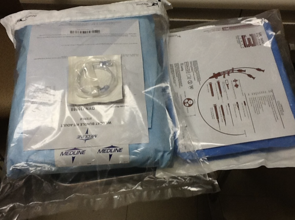
One of the most common and potentially costly complications associated with CVCs is infection. There has been a great deal of focus on this nationwide in order to decrease numbers and prevent infection that can lead to complications, prolonged hospital course, and even death. The most universally preventive measure is aseptic technique.
The use of "packs" and checklists in a controlled environment can help promote and maintain an adequate aseptic technique. Other considerations include prophylactic intravenous antibiotics for high-risk populations (e.g. those on chemotherapy and who are immunocompromised) and “dirty” lines, use of "coated" catheters, selection of insertion site, proper catheter fixation, antiseptic dressings/coverings, proper catheter maintenance, and aseptic technique with use of the catheter.[5:1]
Site selection is very important for CVC placement. It is important for the practitioner to be proficient at multiple sites, but especially at the preferred or primary site. Studies have demonstrated that the subclavian approach is superior to the femoral approach, with femoral approaches being associated with increased risk of colonization and catheter-related sepsis.[6] Studies have not demonstrated a significant difference in the risk of infection associated with the IJ versus SC approach.[7][8][9][10][11] With this in mind, an approach involving the upper body should be the primary consideration, with final choice considering site, contamination, risks, comfort, body habitus, clinical need and comorbid conditions. The femoral approach should only be used as a last resort in emergent situations and should be changed to a more preferred site as soon as possible.
9.3 Benefits of Ultrasound
- Ultrasound guided central line placement is superior to the landmark technique.
- Real time ultrasound should be used throughout central line placement.
- Ultrasound should be used to confirm placement of the wire and catheter.
- Probe marker always to your left.
The use of ultrasound as part of CVC placement has consistently demonstrated superiority to landmark techniques. Static ultrasound has been shown to increase first insertion attempt success rates.[12] Multiple studies have demonstrated that real time ultrasound for the IJ and femoral approaches increases first insertion attempt success rate, decreases access time, increases the likelihood of successful cannulation, and decreases likelihood of arterial puncture.[13][14][15][16][17][18] As for the subclavian approach, research demonstrates that real time ultrasound increases success rates, decreases attempts, decreases time, and decreases arterial puncture.[19][20] However, it should be noted that the subclavian approach is still not routinely performed under ultrasound guidance even by some practitioners who are very adept with ultrasound. Many feel that the clavicle landmark makes the gains achieved by ultrasound guidance less dramatic with this approach. However, the literature does support those gains as real, and ultrasound should be considered.
Ideally, real time ultrasound should be used throughout central line placement. It should first be used to identify and establish the anatomy before site prep. Obstacles, such as a blood clot within the vein of interest, can be identified prior to starting (#Movie 9.1).
Next, US should be used to reorient, view cannulation of the vein, and then to confirm placement of the wire and catheter (#Movie 9.2). The images should be archived for later reference and QA as indicated.
Movie 9.1
IJ Thrombus
##### Movie 9.2 **Wire in IJ** ### Considerations Prior to Procedure Indications: 1) Inability to obtain peripheral IV access. 2) Need for rapid fluid loading or administration of caustic agents. 3) Emergent venous access. 4) CVP monitoring. 5) Transvenous pacer placement. 6) Pulmonary artery catheterization. Contraindications: 1) Absolute: anatomical anomaly, thrombus, or infection at site of insertion. 2) Relative: anticoagulation, bleeding dyscrasia, inability to cooperate, or excessive body habitus. 3) Lack of training or experience.Potential Complications:
- Arterial puncture
- Bleeding/hematoma
- Hemo/pneumothorax
- Thrombosis
- Infection
- Brachial plexus/nerve injury
Equipment (#Gallery 9.1): - Ultrasound machine
- High frequency linear array probe
- Sterile probe cover
- Sterile gel
- CVC kit
- Sterile personal protective equipment
Gallery 9.1
Equipment
[[Screenshot 2025-12-18 at 10.12.22 PM.png]] | Linear probe
[[Screenshot 2025-12-18 at 10.12.37 PM.png]] | Sterile probe cover
[[Screenshot 2025-12-18 at 10.12.53 PM.png]] | Sterile gel
[[Screenshot 2025-12-18 at 10.13.05 PM.png]] | CVC kit
[[Screenshot 2025-12-18 at 10.13.20 PM.png]] | Sterile personal protective equipment
Desired Visualization
Internal Jugular
Visualize with the probe in the transverse position with indicator to the left. There is some slight variability in anatomy, but typically, the vein will appear as a larger, somewhat triangular shaped vessel just anterior and lateral to the artery (#Movie 9.3).
Movie 9.3
Finding the IJ
If the patient is dehydrated or hypotensive, the vein may appear flattened and may even disappear with inspiration. Trendelenburg position and valsalva can assist in visualization (#Movie 9.4 and #Movie 9.5).
Movie 9.4
IJ Valsalva
Movie 9.5
Dehydrated Patient Placed in Trandelenburg
Supraclavicular
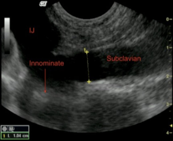
Visualize with the probe in a 45 degree bisection at the angle of the sternocleidomastoid and the clavicle. The subclavian vein can be visualized as it joins the internal jugular vein to form the innominate vein (#Figure 9.1, #Image 9.2 and #Movie 9.6).
Figure 9.1
Supraclavicular Probe Placement
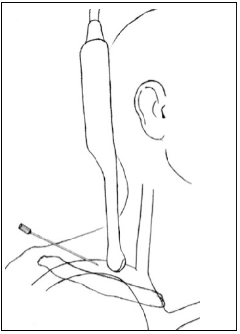
Image 9.2
Supraclavicular View
Movie 9.6
Supraclavicular Placement
Infraclavicular/Subclavian
Visualize with the probe perpendicular to the inferior border of the clavicle at the angle of the clavicle. The vein will appear as a larger, thin walled vessel oriented more cephalad, which will appear on the right of the screen when approaching from the left and will appear on the left of the screen when approaching from the right (#Movie 9.7, see a 3D demonstration).
Movie 9.7
Subclavian Placement
Femoral
Visualize with the probe transverse to the leg at the inguinal ligament. The vein will appear as a larger, thin walled, triangular shaped vessel oriented medially to the artery and the nerve (see a 3D demonstration).
Note: if the patient is dehydrated or hypotensive, the vein may be collapsed, especially if any pressure is placed on the vein. It is important to be familiar with the anatomy in order to understand where the vein should be if it is compressed (#Movie 9.8). Once the artery is visualized, the location of the vein can typically be identified. For approaches other than femoral, the Trendelenburg position and valsalva can assist in visualization of the vein. For the femoral approach, reverse Trendelenburg may assist with expansion of the vein. Removing some of the pressure from the probe may also allow the vein to expand for visualization (Movie 9.8). Understand that if the vein is compressed, it is very easy when inserting the needle to penetrate the anterior and posterior walls of the vessel at the same time without the flash of blood to indicate your location.
Movie 9.8
IJ Compressed with Pressure
9.4 Procedure
- An assistant can be very helpful
- Preparation is key to a successful procedure
Some feel that ultrasound guided CVC placement is best achieved with two people, one to place the line and the other to manage the ultrasound. It is possible to do with one person, but it is more difficult and requires increased coordination and organization, especially for novices. Once one is adept with the ultrasound, then one-person placement can be just as easy and perhaps easier.
Regardless of technique used, it can be very helpful to practice on a simulation device prior to performing on a real patient.[21]
Procedure
- Obtain informed consent from the patient for the procedure and perform "time out" before starting.
- Obtain all supplies necessary and arrange at the bedside in a way in which everything is easily accessible (#Gallery 9.2).
- Position the patient in order to maximize visualization and to aid in access to the site during placement.
- Apply gel to the end of the linear array probe.
- Open the kit and don sterile personal protective equipment.
- With assistance, place sterile sleeve over the ultrasound probe and secure in place with sterile rubber band.
- Place sterile gel on end of sleeved probe.
- Prep and drape the patient in a sterile manner.
- Visualize the vein with the ultrasound to establish landmarks (#Movie 9.3).
- Inject overlying tissue with 1% lidocaine for local anesthesia.
- Re-visualize the vein with the ultrasound.
- Insert the echogenic introducer needle at a 45 degree angle and visualize it as it advances towards the vein. As you approach the wall, you will note depression of the anterior wall of the vessel. Slowly advance until a flash of blood is returned. Upon insertion note the depth of the vessel and correlate with the needle in order to avoid overinsertion of the needle (#Movie 9.9 and #Movie 9.10).
- Once blood is noted in the syringe, remove the syringe while holding the needle in place to confirm venous blood via characteristic maroon, nonpulsatile flow. If there is any question, an ultrasound image in long axis may be helpful to confirm needle position.
- Insert the guide wire through the needle and advance until the approximate length of the catheter is visible. Monitor closely for arrythmias associated with wire induced cardiac irritability. If noted, pull the wire back slowly until ectopy resolves. Having an assistant watch wire insertion in long axis can add yet another confirmation of placement (#Movie 9.11).
- Remove the introducer needle (#Movie 9.12).
- Make a small skin incision over the wire (#Movie 9.13).
- Insert the tissue dilator over the wire and advance until almost to the hub and then remove (#Movie 9.14).
- Since the patient is prepped and draped, you can hold the catheter up to the site to determine how far to advance the catheter. Insert the catheter over the wire, but ensure you maintain control over the wire at all times. Typically, you will have to pull back slightly on the wire to ensure it is visible at the end of the catheter to enable you to hold it while inserting the catheter (#Movie 9.15).
- Advance the catheter until the tip would be approximately at the junction of the superior vena cava and right atrium, as previously measured.
- Secure the catheter into place, insert biodisc, and cover with sterile dressing (Movies 9.15, 9.16, and 9.17).
- Aspirate and flush all ports with sterile saline (Movie 9.18).
- Look at lungs with ultrasound to rule out pneumothorax (see Pneumothorax section in FAST/EFAST chapter).
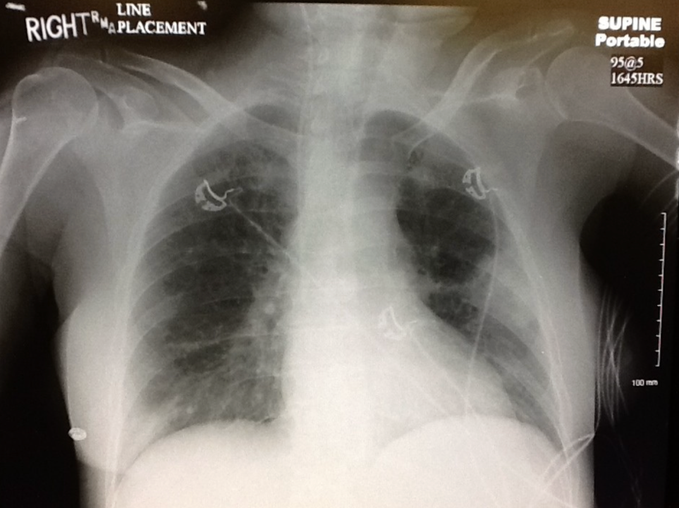
- Obtain a STAT portable chest X-ray to confirm placement of the catheter (Images 9.3-9.4).
Gallery 9.2
CVC Preparation
[[Pasted image 20260308173145.png]] | CVC kit
[[Pasted image 20260308173221.png]] | Linear probe
[[Pasted image 20260308173337.png]] | Personal protective equipment
[[Pasted image 20260308173426.png]] | Sterile probe cover
[[Pasted image 20260308173246.png]] | Sterile gel
Movie 9.9
Place Needle
##### Movie 9.10 **Needle Tip in IJ** ##### Movie 9.11 **Insert Wire** ##### Movie 9.12 **Remove Needle Over Wire** ##### Movie 9.13 **Skin Incision** ##### Movie 9.14 **Dilating** ##### Movie 9.15 **Thread Catheter** ##### Movie 9.16 **Suture in Place** ##### Movie 9.17 **Place Biodisc** ##### Movie 9.18 **Aspirate and Flush**Image 9.3
Stat X-Ray
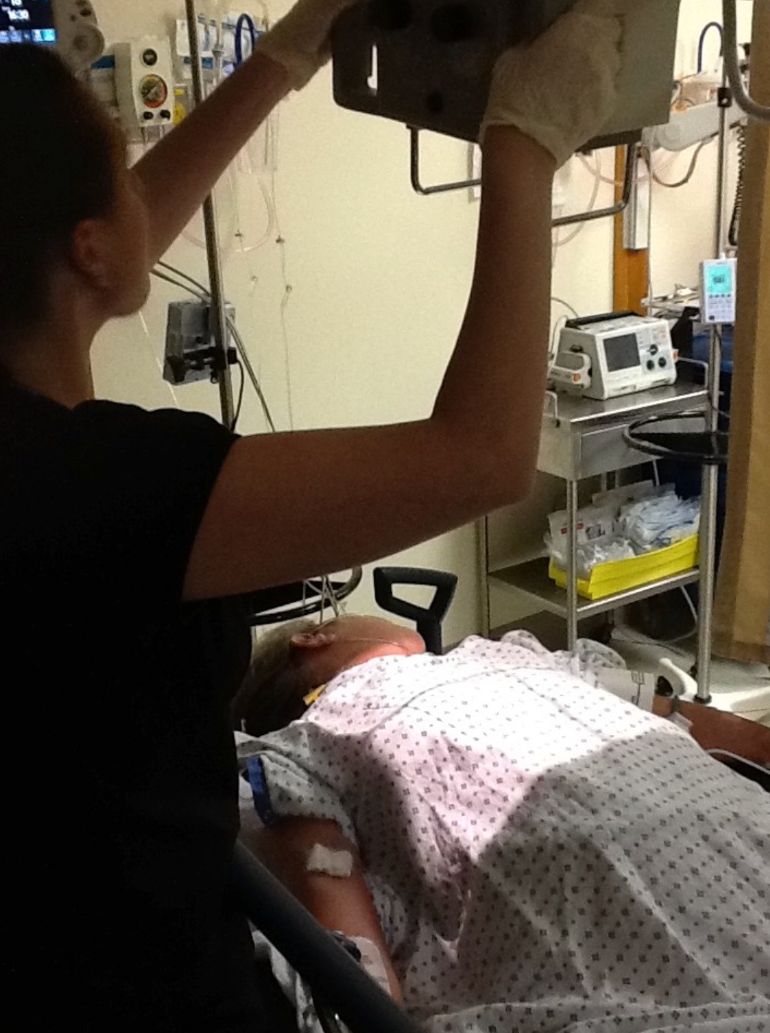
Image 9.4
X-Ray Confirmation
Conclusion
Ultrasound guidance for central line placement is now the standard of care. It has been proven to be both faster and safer than landmark techniques, with fewer complications.
9.5 References
Sznajder J, Zveibil F, Bitterman H, Weiner P, Bursztein S. Central vein catheterization. Failure and complication rates by three percutaneous approaches. Arch Intern Med. Feb 1986; 146(2): 259-61. ↩︎
Jastremski M, Matthias H, Randell P. Femoral venous catheterization during cardiopulmonary resuscitation: a critical appraisal. J Emerg Med. 1984;1(5):387-91. ↩︎
Ullman J, Stoelting R. Internal jugular vein location with the ultrasound Doppler blood flow detector. Anesth Analg. Jan-Feb 1978;57(1):118. ↩︎
Yonei A, Nonoue T, Sari A. Real-time ultrasonic guidance for percutaneous puncture of the internal jugular vein. Anesthesiology. Jun 1986;64(6):830-1. ↩︎
American Society of Anesthesiologists Task Force on Central Venous Access. Practice Guidelines for Central Venous Access. Anesthesiology. 2012; 116:539-73 ↩︎ ↩︎
Merrer J, De Jonghe B, Golliot F, Lefrant J, Raffy B, Barre E, Rigaud J, Casciani D, Misset B, Bosquet C, Outin H, Brun-Buisson C, Nitenburg G. French Catheter Study Group in Intensive Care: Complications of femoral and subclavian venous catheterization in critically ill patients: A randomized controlled trial. JAMA. 2001;286:700-7. ↩︎
Collignon P, Soni N, Pearson I, Sorrell T, Woods P. Sepsis associated with central vein catheters in critically ill patients. Intensive Care Med. 1988;14:227-31. ↩︎
Gil R, Kruse J, Thill-Baharozian M, Carlson R. Triple vs single-lumen central venous catheters: A prospective study in a critically ill population. Arch Intern Med. 1989;149:1139-43. ↩︎
Gowardman J, Robertson I, Parkes S, Rickard C. Influence of insertion site on central venous catheter colonization and bloodstream infection rates. Intensive Care Med. 2008;34:1038-45. ↩︎
Lorente I, Henry C, Martin M, Jiminez A, Mora M. Central venous catheter-related infection in a prospective and observational study of 2,595 catheters. Crit Care. 2005; 9:R631-5. ↩︎
McKinley S, MacKenzie A, Finfer S, Ward R, Penfold J. Incidence and predictors of central venous catheter related infection in intensive care patients. Anesth Intensive Care. 1999;27:164-9. ↩︎
Milling T Jr., Rose J, Briggs W, Birkhahn R, Gaeta T, Bove J, Melniker L. Randomized, controlled clinical trial of point-of-care limited ultrasonography assistance of central venous cannulation: The Third Sonography Outcomes Assessment Program (SOAP-3) Trial. Crit Care Med. 2005;33:1764-9. ↩︎
Karakitsos D, Labropoulos N, De Groot E, et al. Real-time ultrasound guided catheterization of the internal jugular vein: A prospective comparison with the landmark technique in critical care patients. Crit Care. 2006;10:R162. ↩︎
Mallory D, McGee W, Shawker T, Brenner M, Bailey K, Evans R, Parker M, Farmer J, Parillo J. Ultrasound guidance improves the success rate of internal jugular vein cannulation: A prospective, randomized trial. Chest. 1990; 98:157-60. ↩︎
Verghese S, McGill W, Patel R, Sell J, Midgley F, Ruttimann U. Comparison of three techniques for internal jugular vein cannulation in infants. Paediatr Anaesth. 2000; 10:505-11. ↩︎
Verghese S, McGill W, Patel R, Sell J, Midgley F, Ruttimann U. Ultrasound-guided internal jugular venous cannulation in infants: A prospective comparison with the traditional palpation method. Anesthesiology. 1999; 91:71-7. ↩︎
Verghese S, McGill W, Patel R, Sell J, Midgley F, Ruttimann U. Ultrasound-guided internal jugular venous cannulation in infants: a prospective comparison with the traditional palpating method. Anesthesiology. Jul 1999;91(1):71-7. ↩︎
Denys B, Uretsky B, Reddy P. Ultrasound-assisted cannulation of the internal jugular vein. A prospective comparison to the external landmark-guided technique. Circulation. May 1993;87(5):1557-62. ↩︎
Gueltieri E, Deppe S, Sipperly M, Thompson D. Subclavian venous catheterization: Greater success rate for less experienced operators using ultrasound guidance. Crit Care Med. 1995;23:692-7. ↩︎
Fragou M, Gravvanis A, Dimitriou V, et al. Real-time ultrasound-guided subclavian vein cannulation versus the landmark method in critical care patients: A prospective randomized study. Crit Care Med. 2011;39:1607-12. ↩︎
Sekiguchi H, Tokits J, Minami T, Eisen L, Mayo P, Narasimham M. A prerotational, simulation-based workshop improves the safety of central venous catheter insertion: results of a successful internal medicine house staff training program. Chest. Sep 2011; 140(3):652-8. ↩︎